Röntgen hand kind
in der Kinderheilkunde zur Beurteilung von Reifungsverzögerungen von Kindern benutzt.
Für die Orthopädie ist sie zum Beispiel bei der Skoliosebeurteilung von großer Bedeutung. hormonelle Wachstumssteuerung).
Wilhelm Roentgen’s Background
Wilhelm Roentgen (or Röntgen in German) is a name we’re all familiar with, even if our pronunciation of his name might differ from that in his native German.
His pioneering work with X-rays in the late 19th century laid the foundation for modern medical imaging.
Bei Pedbone bieten wir Ihnen eine umfassende Sammlung von Bildern, die die Entwicklung der Knochenkerne in verschiedenen Altersgruppen und Geschlechtern zeigen. It’s a fuzzy image that shows the bones of her hand, and the ring that adorns it.
It still survives today, as a fine-dining restaurant. Some of the beams were penetrating solid objects and exposing sheets of photographic paper, creating shadowy images.
Unsere Bilder sind nach Regionen des Körpers und Altersklassen sortiert und bieten Einblicke in die Entwicklung von Knochen und Gelenken über die Jahre hinweg.
Above, left, an X-ray of his wife's hand; right, one of the first X-rays taken in the UK, of a woman's hand with ring, bracelet and chain of keys circa 1896
Roentgen, a professor of physics at Wurzburg University, in Bavaria, realised that the phenomenon was due to strange beams being emitted by a glass tube he was using during his investigation.
As electricity passed between two electrodes in the tube, the rays had an effect on the photographic plates.
It was his eureka moment.
This would be the only public lecture that Roentgen would ever take on X-rays. They got married six years later, with lots of reservations from Wilhelm’s father, who was unhappy that his son was marrying a woman from a very humble background who was also six years older to him. It is therefore appropriate that we first talk about the (God)father of radiology, and two hand radiographs (with rings) that he famously captured.
Roentgen was born in 1845 in a small German town named Lennep (now part of the city of Remscheid, near Cologne), and moved shortly afterwards to Utrecht in the Netherlands, where he attended school.
Photographic plates near his equipment had started to glow.
Scroll down for video
The first ever X-ray taken of a human (above, on December 22, 1895) was the left hand, complete with wedding and engagement rings, of Anna Bertha Roentgen - the wife of the man who accidentally discovered a form of radiation that would change the face of medicine
Pictured left is Anna Bertha.
His discovery earned him the first Nobel Prize for Physics in 1901. The ability to see inside the human body without invasive procedures transformed medical practices, enabling doctors to diagnose all kinds of diseases. In November 1895, German physicist Wilhelm Conrad Roentgen (right, circa 1896) was conducting an experiment in his lab when he noticed that photographic plates near his equipment had started to glow.
The logo of Café Roentgen features a hand radiograph (complete with the ring), holding a radiopaque cup with “Café Roentgen” written on it in a font reminiscent of the film The Godfather. zur Wachstumsanalyse bei Kindern!
Problemlos durchzuführen ...
... Instead, he selflessly shared his findings with the world, encouraging researchers to better it, ensuring that X-ray technology rapidly spread across the globe without financial barriers.
Roentgen passed away at the age of 77 on February 10, 1923, succumbing to colorectal cancer in Munich, Germany.
Bei Minder- oder Hochwuchs ist durch Bestimmung des Knochenalters im Verhältnis zum tatsächlichen (chronologischen) Alter abschätzbar, ob das Wachstum sich noch im normalen Bereich bewegt oder ob weitere Maßnahmen zu treffen sind (z.B. This image represented a refinement compared to the earlier radiograph of Anna’s hand. Die Kenntnis des noch zu erwartenden Wachstums hat nicht nur psychosoziale Bedeutung, sondern wird z.B.
ein korrigieren des Wirbelsäulenkorsett nicht mehr getragen werden muss. Struggling to find a college that would accept a candidate without a high-school diploma, he eagerly seized an opportunity to study mechanical engineering provided by the Federal Polytechnic Institute in Zurich, and moved to Switzerland. As a professor at the University of Würzburg, he shared academic space with Roentgen, his close friend and colleague.
On 23 January 1896, he was in the audience for a lecture by Roentgen on his recent discovery, who had been invited by the Physical-Medical Society of Würzburg for a talk.
Man kann also anhand der Endgrößenbestimmung entscheiden, wann z.B.
Roentgen died in Munich in 1923, aged 77, from bowel cancer. The fact that we are discussing his legacy on Café Roentgen, a platform named in his honour, is a testament to the enduring impact of his work.
With their ability to penetrate solid objects, Roentgen's rays would go on to have a wide range of uses, notably in medicine, archaeology and astronomy.
Despite his success in the field of X-rays, he abandoned his work on them a year after their discovery, and instead focused on examining crystals.
Within a year of the beam's discovery, the world's first radiology department opened, in Glasgow Royal Infirmary.
Dies ist von Bedeutung, da es Kinder gibt, die bereits im Alter von 12 ausgewachsen sind, während andere mit 18 noch wachsen. Countless radiology professionals have seen this image, making it perhaps the most defining emblem of the field.
A close-up of the Roentgen tube used for X-ray production - named after the physicist. If there is any actual physical establishment in the world that could potentially call itself “Café Roentgen”, it’s this one.
Although some labelled the beams Roentgen rays, he preferred the term X-rays
Roentgen, a professor of physics at Wurzburg University, in Bavaria, realised the phenomenon was due to strange beams being emitted by a glass tube he was using during his investigation. Anhand dieses Röntgenbildes kann das so ermittelte “Knochenalter” mit dem tatsächlichen sogenannten chronologischen Alter verglichen werden, daraus wird mit Hilfe eines Computerprogrammes die Endgröße des Kindes ermittelt.
Wirkungsweise und Methode
Während der Entwicklung vom Säugling zum Erwachsenen zeigen sich charakteristische Veränderungen an den Wachstumsfugen, die einer genau definierten biologischen Reife zugeordnet werden.
Amazed by what he saw, von Kölliker suggested the unknown rays be called “Roentgen Rays”. This radiograph marks a seminal moment in medical history, and Anna’s contribution to the world of radiology remains invaluable.
But there is another radiograph captured by Roentgen that is often confused with this one, and that too has an interesting story associated with it.
Born in 1817, Albert von Kölliker was a Swiss anatomist best known for discovering the mitochondrion – the powerhouse of the cell.
His enduring legacy lives on, not only in the multitude of lives saved through early and precise diagnoses facilitated by various imaging techniques but also in his spirit of scientific generosity – a spirit that continues to inspire researchers to this day.
In closing, I’ll leave you with the following link of the café where Wilhelm Roentgen met his wife, Anna Bertha.
He was also the first person to demonstrate that smooth muscle is made of many small nucleated muscle cells, and that nerve fibres are continuous with nerve cells. rechtzeitig und präzise!
... Roentgen’s pioneering work with X-rays was quickly recognized, earning him the Nobel Prize in Physics in 1901 (the first year the Nobel Prizes were given out), a testament to the profound impact of his discovery on the scientific community.
Wir laden Sie ein, in unsere Welt der kindlichen Radiologie einzutauchen und von unserem Angebot zu profitieren.
Update:
Neue Bilder sind:
- Becken männlich 11 Jahre
- Becken männlich 14 Jahre
- Hand männlich und weiblich 1-20 Jahre
When she saw the silhouette of the skeleton of her hand, she remarked, “I have seen my own death.”
Wilhelm met his wife Anna in 1866 in Zurich, at “Zunfthaus Zum Grunen Glas” (The Green Glass Guildhouse), a café run by Anna’s father.
Kinderbehandlung / Endgrößenbestimmung
Erkennung von Wachstumsstörungen und Einschätzung der Reife
Die computergestützte Größenbestimmung Ihres Kindes zur Berechnung der zukünftigen Erwachsenengröße dient zur
...
The audience agreed in a standing ovation.
Roentgen’s radiograph of von Kölliker’s hand marked a significant advancement in the nascent field of radiography. In a Science Museum poll in 2009, the X-ray was voted by the British public as the most important modern discovery
Share or comment on this article: Roentgen's first human X-ray of his wife's hand in 1895
The comments below have not been moderated.
The views expressed in the contents above are those of our users and do not necessarily reflect the views of MailOnline.
He eventually graduated with a PhD from the University of Zurich and moved back to his home country – as a faculty member first at the University of Strasbourg, followed by the universities at Giessen, Würzburg and Munich.
In 1895, while working at the University of Würzburg, Roentgen was studying various kinds of vacuum tubes, focusing on what happened when electrical discharges travelled through these tubes.
The radiograph of von Kölliker’s hand not only demonstrated Roentgen’s mastery of the X-ray technique but also demonstrated the scope for future advancements in the field of medical imaging.
Wilhelm Roentgen’s discovery of X-rays in 1895 truly revolutionized the landscape of medicine, opening a realm of diagnosis and exploration previously unimaginable.
During the lecture, Roentgen called von Kölliker to the stage as a volunteer and captured a radiograph of his hand.
The 50-year-old had stumbled upon a new kind of ray: X-radiation, which is composed of X-rays and is a form of high-frequency electromagnetic radiation.
Roentgen referred to the radiation as 'x', which is used in mathematics to represent an unknown quantity.
He then began making X-ray images - or radiographs - of inanimate objects such as weights and a piece of metal.
Naming them “X-rays” (with “X” representing the unknown), Roentgen dedicated himself to their study, and in this process captured the first radiographic image — a flickering silhouette of his own skeleton, an image that is unfortunately lost.
We are no longer accepting comments on this article.
Willkommen zu unserem Angebot zur kindlichen Radiologie!
neues Diagnostikverfahren!
Anwendungsbereiche (Indikationen)
bei kindlichen Skoliosen (Wirbelsäulenverkrümmungen)
bei Wachstumsverzögerung (in der Klasse sind alle deutlich größer)
bei übermäßigem Wachstum (der / die Größte in der Klasse)
bei deutlichen Beinlängenunterschieden
bei Verkrümmungen der Beine (X- oder O-Bein)
bei bestimmten Stoffwechselerkrankungen
Wie wird sie bestimmt?
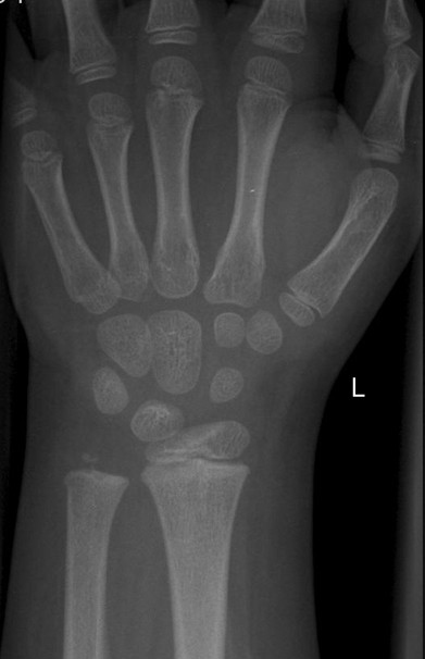
Zur Beurteilung der Knochenreife wird wegen der zahlreichen Wachstumsfugen das Röntgenbild der linken Hand herangezogen.
In a Science Museum poll in 2009, the X-ray was voted by the British public as the most important modern discovery.
The antibiotic agent penicillin came second followed by the DNA double helix.
Physicians diagnose pulmonary tuberculosis - with the aid of X-rays - circa 1900. To his astonishment, he observed a faint shimmering image of a nearby bench each time he discharged the tube.
Before his breakthrough, Roentgen had been studying the effects of passing an electrical current through gases at low pressure. Within a year of the beam's discovery, the world's first radiology department opened, in Glasgow Royal Infirmary
Roentgen (statue, above, on the Potsdam Bridge in Berlin) died in Munich in 1923, aged 77, from bowel cancer.
The father of radiology, as he is rightfully known, has truly left an indelible legacy in the world of science and medicine.
Some of the rays were penetrating solid objects and exposing sheets of photographic paper, creating shadowy images. In the weeks following his discovery, Roentgen diligently refined and fine-tuned the technical aspects of the imaging process. She suffered from renal issues for years and died at the age of eighty years.
Anna’s hand radiograph, a part of Roentgen’s personal collection, is truly iconic, to say the least.
This would lead to his eureka moment and the discovery of X-rays - the 'x' indicating that they were of an unknown type
Then aged 50, Roentgen had discovered a new kind of ray: X-radiation, which is composed of X-rays and is a form of high-frequency electromagnetic radiation. His meticulous adjustments led to significantly enhanced clarity, allowing him to capture the more intricate details of the anatomy of von Kölliker’s hand.
He was unfortunately expelled from high school when one of his teachers intercepted a caricature of one of the other teachers that he reportedly did not draw.

His impact on the world of biology is truly immense. In early November, while working on a Lenard’s tube (a kind of vacuum tube made of thin glass), he noticed that some invisible rays caused fluorescence on a cardboard screen coated with barium platinocyanide, placed near the aluminium window. This intrigued him to consider whether the Crookes-Hittorf tube, with thicker glass walls, could produce a similar effect.
On the fateful afternoon of 8 November 1895, Roentgen connected the Crookes-Hittorf tube to a pair of electrodes and dimmed the room.
Wir unterscheiden auch zwischen den Geschlechtern, und falls das Geschlecht des Patienten nicht bekannt ist, wird die Aufnahme unter "unbekannt" abgelegt.
Unsere Sammlung ist eine wertvolle Ressource für alle, die in der Radiologie von Kindern tätig sind oder ihr Wissen in diesem Bereich vertiefen möchten.
Around six weeks after his groundbreaking discovery, Roentgen captured another radiograph – this time of the hand of his wife, Anna Bertha. He noticed that as electricity passed between two electrodes in the tube, the rays had an effect on the photographic plates
Equipment, including a Roentgen X-ray machine, used by German surgeons in an open-air surgery tent, during the First World War. With their ability to penetrate solid objects, Roentgen's rays would go on to have a wide range of uses, notably in medicine, archaeology and astronomy
An illustration of Roentgen at work in his lab.
Wir haben diese Sammlung sorgfältig zusammengestellt, um medizinischen Fachkräften und Radiologen bei der Befundung von Kinderbildern zu helfen. This showcased the potential of X-ray technology for medical diagnostics, highlighting specific bone structures and nuances previously invisible to the human eye. His demise marked a profound loss for the medical community.
Remarkably, Roentgen chose not to patent his radiographic technique. They never had any children of their own, but adopted a little girl named Josephine, the daughter of Anna’s brother – after his unfortunate demise. He enjoys writing about radiology, history and culture. Despite his success in the field of X-rays, he abandoned his work on them a year after their discovery - to focus on examining crystals
On January 5, 1896, his findings - which included the picture of the bones of his wife's hand - were published to wide acclaim.
You can check out his blog at https://syndrome.home.blog/ and you can reach out to him at anmoldhawan@gmail.com.
Related
The doctor will see through you now: How a German physicist's accidental discovery led to the world's first human X-ray (of his wife's hand) and changed the face of medical diagnosis
To the casual observer, the X-ray of a hand, seen below, appears unremarkable.
However, it is in fact the world's first X-ray taken of a human, in December 1895 - and belongs to the wife of the man who accidentally changed the face of medical diagnosis.
Such was the shock Anna Bertha Roentgen felt upon seeing the skeletal picture of her left hand, complete with wedding and engagement rings, that she exclaimed: 'I have seen my death.'
On November 8 that year, her husband, German physicist Wilhelm Conrad Roentgen, had been conducting an experiment in his lab - the effects of passing an electrical current through gases at low pressure - when something caught his eye.
Beyond the accolades and the fame that he received, his story represents the essence of curiosity, dedication, and the relentless pursuit of knowledge that continues to inspire people around the world.
Roentgen’s legacy is truly remarkable, stretching across disciplines, generations and geographical boundaries.
Es ist wichtig zu wissen, wie viel das betroffene Kind noch wächst, da die Zunahme der Wirbelsäulenverbiegung (Skoliose) nur während des Wachstums zu befürchten ist. Do visit it if you ever find yourself in Zurich – I know I will some day!
– Dr Anmol Dhawan
Dr Anmol Dhawan is a radiologist working as a senior resident at Bharati Hospital, Pune.